Yearly Archives: 2021

Utility Of Urine Examination

The examination of urine for disease diagnosis goes back thousands of years! Ancient Babylonian and Sumerian physicians first inscribed their evaluations of urine into clay tablets as early as 4,000 B.C.

Later, in ancient Greece, Hippocrates, often called the father of Western medicine, expanded on urine’s importance: “No other organ system or organ of the human body provides so much information by its excretion as does the urinary system,” he wrote.Medieval doctors associated nearly every known disease with urinary characteristics and some would diagnose patients without even meeting them!

This simple yet valuable exam is done on a routine basis in all laboratories from the smallest to the biggest! The exam not only helps us with diagnosis of urinary tract related diseases but also diseases of other systems including metabolic disorders like diabetes.

Many disorders may be detected in their early stages by identifying substances that are not normally present in the urine and/or by measuring abnormal levels of certain substances. They may be present because these substances are eliminated from our bodies through urine.

urinary-system

Urine is produced by the kidneys, two fist-sized organs located on either side of the spine at the bottom of the ribcage. The kidneys filter wastes out of the blood, help regulate the amount of water in the body, and conserve proteins, electrolytes, and other compounds that the body can reuse. Anything that is not needed is eliminated in the urine, traveling from the kidneys through ureters to the bladder and then through the urethra and out of the body.

Urine is an unstable fluid, and changes to its composition begin to take place as soon as it is voided. As such, collection, storage, and handling are important issues in maintaining the integrity of this specimen.Hence, it is best to collect the urine sample in the laboratory itself. A urine sample will only be useful for a urinalysis if taken to the laboratory for processing within a short period.However, if the urine is collected at home or in the doctor’s clinic, if it will be longer than an hour between collection and transport time, then the urine should be refrigerated or a preservative may be added.

What is being tested?

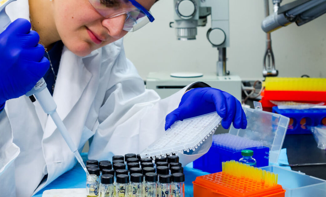
A urinalysis is a group of physical, chemical, and microscopic tests. The tests detect and/or measure several substances in the urine, such as byproducts of normal and abnormal metabolism, cells, cellular fragments, and bacteria.

In the laboratory, urine can be characterized by physical appearance, chemical composition, and microscopically. Physical examination of urine includes description of color, odor, clarity, volume, and specific gravity. Chemical examination of urine includes the identification of protein, blood cells, glucose, pH, bilirubin, urobilinogen, ketone bodies, nitrites.

Finally, microscopic examination entails the detection of crystals, cells, casts, and microorganisms.

All the findings that are generated by this simple exam can give valuable information to the treating physician to guide and cure the patient.

How is the sample collected for testing?

One to two ounces of urine is collected in a clean container. A sufficient sample is required for accurate results.

Urine for a urinalysis can be collected at any time. In some cases, a first morning sample may be requested because it is more concentrated and more likely to detect abnormalities.

Sometimes, you may be asked to collect a “clean-catch” urine sample. For this, it is important to clean the genital area before collecting the urine. Bacteria and cells from the surrounding skin can contaminate the sample and interfere with the interpretation of test results. With women, menstrual blood and vaginal secretions can also be a source of contamination. The genital area should be cleaned with warm water before collection. Start to urinate, let some urine fall into the toilet, then collect one to two ounces of urine in the container provided, then void the rest into the toilet.

When is examination of urine ordered?

A urinalysis may sometimes be ordered when a person has a routine wellness exam, is admitted to the hospital, or will undergo surgery, or when a woman haspregnancycheckup.

A urinalysis will likely be ordered when a person sees a doctor, complaining of symptoms of a urinary tract infection or other urinary system problem, such as kidney disease.Some signs and symptoms may include:

  • Abdominal pain
  • Back pain
  • Painful or frequent urination
  • Blood in the urine
  • Testing may also be ordered at regular intervals when monitoring certain conditions like diabetes and drugs.

A simple urine exam can go a long way in diagnosing and help in treating or managing problems not only relating to the urinary system but other systems too!

Hepatitis: All you need to know about this liver disease

Liver

Liver is the largest organ in the body and is located in the right upper quadrant of the abdomen &accounts for 1.5 to 2.5 % of lean body mass. Diseases of liver present clinically in a few distinct patterns and are usually classified as hepatocellular diseases, cholestaticdiseases or mixed.

Hepatitis is inflammation of the liver parenchyma. The condition may be clinically in-apparentand self limiting, or may be chronic progressing to fibrosis or scarring of liver, eventually resulting in cirrhosis and liver cancers.

Causes of Hepatitis :

  • Viral infections
  • Toxins& drug induced
  • Autoimmune disorders.

Viral hepatitis :

Viral hepatitis is asystemic infection affecting the liver predominantly. There are five different types of Hepatitis viruses, identified by different letters A,B,C,D and E. While all cause liver disease they vary in important ways. In particular, Hepatitis B and hepatitis C lead to chronic disease in hundreds & millions of people and are among the most common causes of liver cirrhosis and cancer.

Most people do not experience any symptoms during the acute infection phase. However, some people have acute illness with symptoms that last several weeks, including yellowing of the skin and eyes (jaundice), dark urine, extreme fatigue, nausea, vomiting and abdominal pain. Jaundice is the hallmark of liver disease and perhaps the most reliable marker of severity.A small subset of persons with acute hepatitis can develop acute liver failure, which can lead to death.

In some people, hepatitis can also cause a chronic liver infection that can later develop into cirrhosis (a scarring of the liver) or liver cancer.

Hepatitis A and E are typically caused by ingestion of contaminated food or water. Hepatitis B, C and D usually occur as a result of parenteral contact with infected body fluids. Common modes of transmission for these viruses include receipt of contaminated blood or blood products, invasive medical procedures using contaminated equipment and for hepatitis B transmission from mother to baby at birth, from family member to child, and also by sexual contact.

Hepatitis B is a potentially life-threatening liver infection caused by the hepatitis B virus (HBV) that can cause chronic infection and puts people at high risk of death from cirrhosis and liver cancer.

A vaccine against hepatitis B has been available. The vaccine is 95% effective in preventing infection and the development of chronic disease and liver cancer due to hepatitis B.

Toxic & drug induced hepatitis:

  • Among patients with acute liver failure, drug induced hepatitis is the common cause.Hepatotoxicity may be direct or idiosyncratic. Most commonly used drugs that cause hepatitis includes anticonvulsants like Phenytoin, Phenobarbitone, antifungal agents like ketaconazole, Anti TB drugs like Isoniazid, rifampicin.
  • Direct hepatotoxicity is dose dependent, predictable and has a short interval between exposure and liver injury.Idiosyncratic reactions are unpredictable, may occur at anytime during or shortly after exposure to the drug
  • Treatment of drug induced hepatitis is largely supportive. Withdrawal of the suspected agent is indicated at the first sign of an adverse reaction.

Autoimmune hepatitis:

Autoimmune hepatitis is a chronic disorder characterized by hepatocellular necrosis, usually with fibrosis progressing to cirrhosis and liver failure.Evidence suggests that the progressive liver injury in patients with autoimmune hepatitis is a result of cell mediated immunological attack directed against liver cells. Most often predisposition to autoimmunity is inherited. Mainstay of management of autoimmune hepatitis is glucocorticoid therapy.

Important diagnostic tests in liver diseases:

Typical battery of tests used for initial assessment of liver diseases includes measurements of

  • Serum aminotransferases
  • Alkaline phosphatase
  • Bilirubin levels
  • Prothrombin time
  • Hepatitis serology : HbsAg, Anti HBc IgM, HBeAg, Anti HCV, Anti HAV IgM
  • Antinuclear antibody (ANA),Antimitochondrialantibody (AMA), P-ANCA
  • Drug history
  • Imaging studies : Ultrasound, CT and MRI scans.
  • Liver biopsy remains the standard criterion, particularly in evaluation of patients with chronic liver disease

Management of liver diseases includes advice of alcohol use, medications, vaccinations and surveillance for complications. Abstinence from alcohol should be encouraged for all patients with alcohol related liver disease.

All patients with risk factors for liver disease should be vaccinated against Hepatitis A and Hepatitis B. Cirrhosis of liver warrants screening & long term surveillance for hepatocellular carcinoma.

Thyroid: The issues and therapies

  • Thyroid is the largest endocrine gland in the body. It is located in the neck, wrapped around the windpipe and is made of follicles that produces and stores thyroid hormones:thyroxine (T4) and triiodothyronine (T3). These hormones play a critical role in many fundamental processes including cell differentiation, growth, thermogenic and metabolic functions.
  • Production of thyroid hormones is regulated by a complex interplay involving thyroidstimulating hormone (TSH) secreted by the pituitary gland, and thyrotropin releasing hormone (TRH) secreted by the hypothalamus, as well as other factors.

Thyroid disorders:
Disorders of thyroid gland can result in glandular destruction and hormone deficiency (hypothyroidism) or overproduction of thyroid hormones (thyrotoxicosis). In addition, benign nodules and various forms of cancers are relatively common in the thyroid gland.

Hypothyroidism :
Insufficient production of thyroid hormones results in hypothyroidism. Iodine deficiency remains a common cause of hypothyroidism worldwide. In areas of iodine sufficiency autoimmune thyroiditis ( Hashimoto’s thyroiditis) and iatrogenic causes are most common.

Autoimmune thyroiditis :
As with most autoimmune disorders, susceptibility is determined by a combination of genetic & environmental factors. Patients with Hypothyroidism usually present with Tiredness, dry skin, feeling cold, weight gain, poor concentration &, menstrual disorders. Fertility is reduced and incidence of miscarriage is increased.

Neonatal hypothyroidism is due to thyroid gland dysgenesis in 80 -85%. Many present clinically with prolonged jaundice, feeding problems, hypotonia, enlarged tongue. Permanent neurological damage results if treatment is delayed.

Hypothyroidism is generally treated with daily replacement dose of levothyroxine.

Thyrotoxicosis and Hyperthyroidism:
Thyrotoxicosis is a state of thyroid hormone excess. Most common causes of thyrotoxicosis are hyperthyroidism caused by Grave’s disease, toxic multinodular goiter and toxic adenomas. Unexplained weight loss, irritability, heat intolerance and sweating, palpitations, menstrual disorders, fatigue and weakness may be symptoms associated with thyrotoxicosis. The clinical presentation depends on the severity of thyrotoxicosis, duration of disease and patient’s age.

Subacute thyroiditis or viral thyroiditis :
Many viruses have been implicated in viral thyroiditis including mumps, coxsackie, influenza, and adenoviruses. Generally, patients presents with a painful and enlarged thyroid sometimes accompanied by fever. There may be features of thyrotoxicosis or hypothyroidism depending on the phase of the illness. Complete resolution is the usual outcome.

Goiters and nodular thyroid disease:
Goiter refers to an enlarged thyroid gland. Iodine deficiency, autoimmune disease and nodular thyroid disease can all result in goiter. In iodine deficient areas, thyroid enlargement reflects a compensatory effort to produce sufficient thyroid hormone. Endemic goiter is also caused by exposure to environmental goitrogens such as cassava roots, vegetables like cabbage and cauliflower. Most goiters are asymptomatic. However, thyroid function tests should be performed in all patients with goiter to exclude hypothyroidism or thyrotoxicosis.

Thyroid cancer:
Thyroid cancer is twice more common in women, but male gender is associated with worse prognosis. Risk factors for thyroid cancers include exposure to ionizing radiation, new or enlarging neck mass, family history of thyroid cancer.

Thyroid carcinoma is the most common cancer of the endocrine system. Differentiated tumors such as papillary thyroid cancers or follicular thyroid cancer are often curable.

Laboratory evaluation of thyroid disorders:

Enhanced sensitivity and specificity of TSH assays have greatly improved laboratory assessment of thyroid function

Thyroid stimulating hormone (TSH), Free T4 and Free T3 levels:
Logical approach to thyroid testing is to first determine if TSH is normal, suppressed or elevated. The finding of an abnormal TSH must be followed by measurements of circulating thyroid hormone levels to confirm the diagnosis of hypothyroidism or hyperthyroidism. T4 and T3 are highly protein bound, and numerous factors influence protein bounding. It is therefore better to measure free, unbound thyroid hormones.

Tests to determine etiology of thyroid dysfunction:

Autoimmune thyroid dysfunction is easily determined by measuring circulating antibodies against Thyroid peroxidase (TPO) and thyroglobulin (Tg). Almost all patients with autoimmune hypothyroidism and 80 % of patients with Grave’s disease have high levels of TPO antibodies.

Ultrasound scanand USG guided FNA cytology:

Ultrasound scan is valuable is diagnosis and evaluation of nodular thyroid disease. Certain sonographic patterns are highly suggestive of malignancy ( hypoechoic solid nodules, microcalcifications) where as other features correlate with benignity. Ultrasound guided FNA cytology, imaging of the cervical lymph nodes are indispensable is evaluation of patients of thyroid cancers.

Radioiodide uptake and thyroid scanning:

Thyroid scintigraphy should be used in evaluation of patients with thyroid nodules, especially when TSH levels are subnormal.

Screening every 5 years by measuring serum TSH is recommended for all men ≥ 65 and for all women ≥ 35. Screening is also recommended for all newborns and for pregnant women. For those with risk factors for thyroid disease, the serum TSH should be checked more often. Screening for hypothyroidism is as cost effective as screening for hypertension, hypercholesterolemia, and breast cancer. This single test is highly sensitive and specific in diagnosing or excluding two prevalent and serious disorders (hypothyroidism and hyperthyroidism), both of which can be treated effectively. Because of the high incidence of hypothyroidism in the elderly, screening on an annual basis is reasonable for those > age 70.

X
Open chat